DMSE researchers develop a low-cost technique to get lithium out of rocks
Demand for lithium has surged in recent years as lithium-ion batteries power increasingly more of our world. And yet, even as places like the U.S., Europe, and Australia have abundant lithium resources within their borders, China dominates global lithium refining. The biggest hurdle to tapping into the U.S. and Australia’s lithium is getting it out of hard rock minerals in a form that is useful.
Extracting lithium from hard rock today is an energy- and waste-intensive process that is often far more expensive than getting lithium from brine water, which also has major environmental drawbacks. Currently, lithium hard rock extraction involves baking the rock at over 1,000 Celsius and chemically leaching it to extract lithium. The rest of the rock is discarded.
Now, a team of researchers from MIT and elsewhere has developed a low-temperature process for extracting battery-grade lithium from the most common type of lithium-bearing mineral. The process uses a liquid reagent to dissolve the rock into the useful forms of its constituent parts: not just battery-ready lithium salts, but also smelter-grade alumina and cement-ready silica. After the minerals are extracted, the solvent and reagent can be recovered and used again so waste levels approach zero.
The researchers estimate the closed-loop process is half the cost of traditional lithium hard rock extraction and could make it cost-competitive with extracting lithium from brine water.
A paper describing the process was published today in Science. The researchers have already begun commercializing the technology through an MIT spinout, Rock Zero.
“By 2040, we need to quadruple production of lithium globally, which amounts to hundreds of new lithium producing assets,” says author Camden Hunt, a former project manager in MIT’s Center for Electrification and Decarbonization of Industry. “Hard rock is abundant; you can find it everywhere. But most hard rock refining is done in China. Our central thesis is if you can find an easier way to crack the rock, get lithium out, and make battery-grade lithium salts, you can change the lithium market. It aligns with the recent push to onshore production of critical minerals in the U.S.”
Joining Hunt on the paper are former MIT postdoc Benjamin Mowbray; PhD candidate Kalyn Fuelling; MIT undergraduate Jacqueline Prawira; Khashayar Jafari, a former senior research scientist at the MIT green cement spinout Sublime Systems; and Yet-Ming Chiang, MIT’s Kyocera Professor of Materials Science and Engineering.
From bathrooms to batteries
The research has its roots in a bathroom renovation. About 25 years ago, as Chiang made a trip to a hardware store to look for something that would turn clear glass blocks translucent, he stumbled on a glass etching cream that works by “eating away” at the surface of the glass. The active ingredient turned out to be ammonium fluoride.
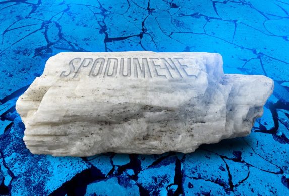
More recently, as Chiang was brainstorming ways to chemically break apart the most abundant lithium-bearing mineral, spodumene, he thought back to that etching cream. Spodumene, like glass, consists mostly of silica. Conventional chemistry-based methods for extracting metals from ores preferentially dissolve more reactive elements and leave behind a silica-enriched residue because of the strength of silicon-oxygen bonds. By designing their process to use a mixture of water and ammonium fluoride, the researchers are able to dissolve silica first, reversing the process.
The researchers showed they could dissolve spodumene rock at room temperature, which represented a breakthrough over traditional processes requiring extreme heat. But it was still only the first step to a closed-loop system that produced useful materials.
“Dissolving silica is the hard part in mining,” Mowbray says. “The next question was how do we apply it to impactful mineral processing problems?”
The mineral spodumene is mainly made up of three components: lithium, aluminum, and silica. Mowbray and Hunt, who both have their PhDs in chemistry, began exploring ways to refine those components separately after they were broken apart in the ammonium fluoride solution.
First, the researchers isolated lithium fluoride, a useful input for common electrolyte materials used in batteries.
Chiang, who has founded several battery companies over his multi-decade career at MIT, next asked the research team if they could isolate lithium hydroxide and lithium carbonate, two lithium salts useful for making battery cathodes. The researchers went back to the lab and found they could make both by developing new processes, some of which involved adding carbon dioxide or sodium carbonate. Chiang tasked the research team with a similar challenge for the aluminum part of the rock, which was isolated using a high-temperature separation technique, and then silica, which was isolated by precipitation.
“First our goal was to produce these products, then there were additional steps of characterizing their purity and properties and making sure our products met the specifications for target markets,” Mowbray explains.
“For the lithium salts, we identified the purity specifications for battery-grade lithium carbonate, the most widely used lithium salt. For the silica, we wanted it to be used as a cement additive, so we did cement reactivity tests and eventually created cubes of cement from it for strength testing using industrial methods. For aluminum, we targeted smelter-grade aluminum. If any product didn’t meet the target specs, you’d end up with a waste stream.”
The researchers then developed a process to reuse the ammonium fluoride and water that starts the reaction.
“We’re able to dissolve the rock with the spodumene in it, and that liberates all the elements, including the aluminum and lithium,” Chiang says. “The silica is in the solution, but on the way to making ammonium fluoride, ammonia gas also comes off. If that ammonia gas is then reapplied, it precipitates the silica again. That sequence gives us back the starting ammonium fluoride. That’s why it’s a circular process.”
The researchers successfully processed 17 different spodumene rock sources, showing its widespread applicability using rocks around the world.
“You’ve heard of nose-to-tail eating?” Chiang says. “We refer to this as nose-to-tail mining. Our researchers came to MIT to look for impactful problems to work on in sustainability. With their skill sets, it was just a matter of setting them loose on this problem. We went through all these steps, and for each one, I’d just say, ‘Can you do this next step?’ And a week or two later they’d say, ‘Okay, we’ve shown we can do that.’ That’s how this entire process got built.”
Scaling the process
Chiang further challenged his research team to evaluate the commercial feasibility of their new system.
“Once we had these core operations worked out, Yet encouraged us to do some math,” Mowbray explains. “Is there enough spodumene in the world to supply 100 terrawatt-hours of battery production? The follow up was: If you supply all the world’s batteries with this process, what are the volumes of the co-products? Do they match global commodity markets? Then we started looking at the cost of the reagents, the cost of the energy, equipment. We started gaining conviction that this could have a big impact.”
The work has special significance for Mowbray, who grew up in a historic mining town in rural British Columbia.
The researchers worked with MIT’s Technology Licensing Office to spin out their company, Rock Zero, which is now located at The Engine and scaling up the system.
“We believe this approach is the lowest-energy, lowest-cost way of getting lithium not only out of hard rock, but period,” Chiang says. “That’s what’s motivating us to scale this. It will enable the energy transition through batteries that use lithium. This was one of the goals of The Climate Project at MIT — to work on projects that, within a short number of years, could transition from the lab to commercialization and impact.”
The work was supported, in part, by the Department of Energy Advanced Research Projects Agency-Energy (ARPA-E), the MIT Climate Grant Challenges program, and the National Science Foundation. The work made use of MIT.nano facilities.
Related Stories