Microscope Image Contest
Microscope imagery is central to the Breakerspace’s educational mission. The Microscope Image Contest showcases the creativity and technical skill of MIT undergraduates as they explore the microscopic world.
Winter 2026
The second edition of the competition drew a wide range of images, from split hairs to fossilized remains of algae.
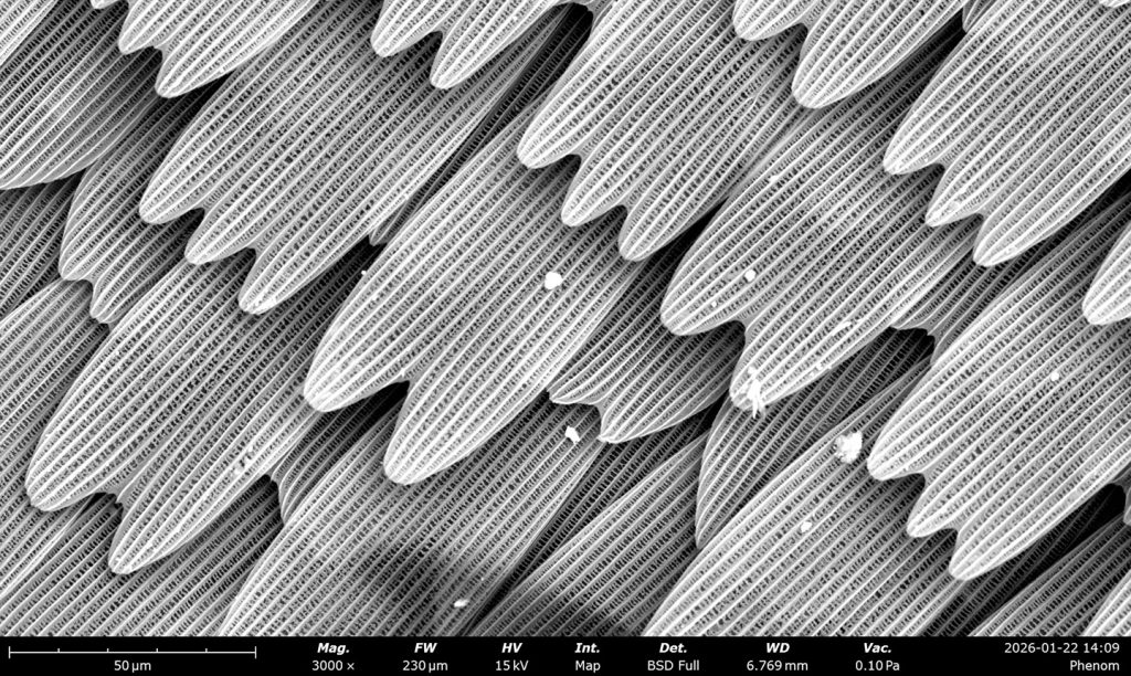
Shingle-like scales on a butterfly wing. The specimen preserves scales with patterns reminiscent of shark denticles, highlighting structural similarities across different species.
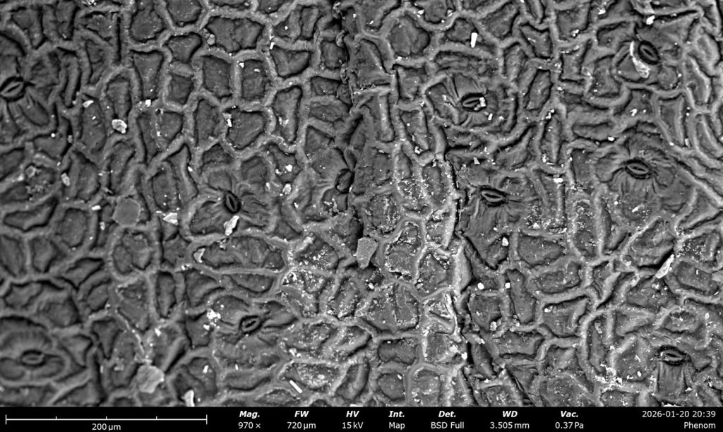
The surface of a succulent leaf, revealing sunken stomata that help the plant thrive with little water, along with interlocking pavement cells. The image highlights how microscopic structure supports function and survival.
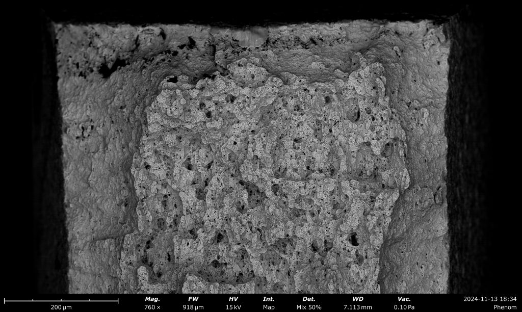
Activated charcoal used for medicinal purposes, typically made from plant sources.
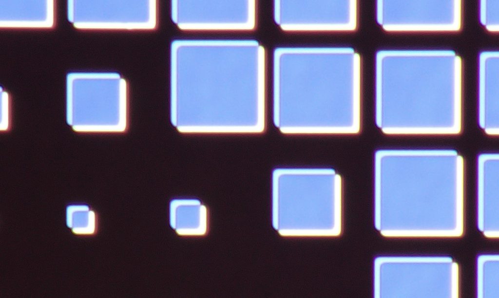
A silicon wafer patterned with aluminum. Regular geometric shapes feel almost computer-generated. The image reflects scientific ingenuity in our ability to fabricate and visualize flawless structures at tiny scales.
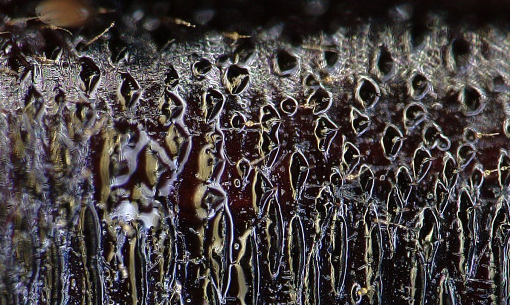
The underside of a damselfly abdomen, revealing icicle-like structures on the exoskeleton.
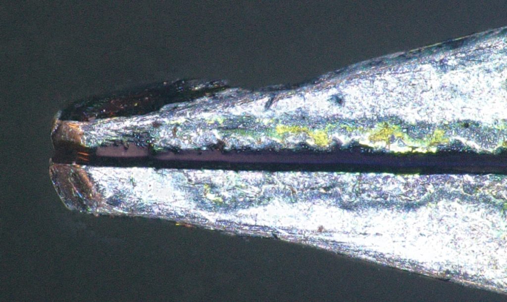
The nib of an Esterbrook J Series fountain pen, produced between 1948 and 1957. The nib slit shows erosion from more than 65 years of wear.
Fall 2024
Below are the winning images from the inaugural competition, which drew dozens of stunning images.
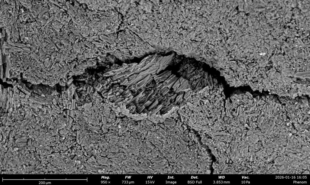
Fractograph of a titanium alloy showing a partially brittle, partially ductile fracture caused by electrochemical hydrogen embrittlement. Hydrogen diffuses inward from the edges, leaving a rectangular patch of ductile metal at the center.
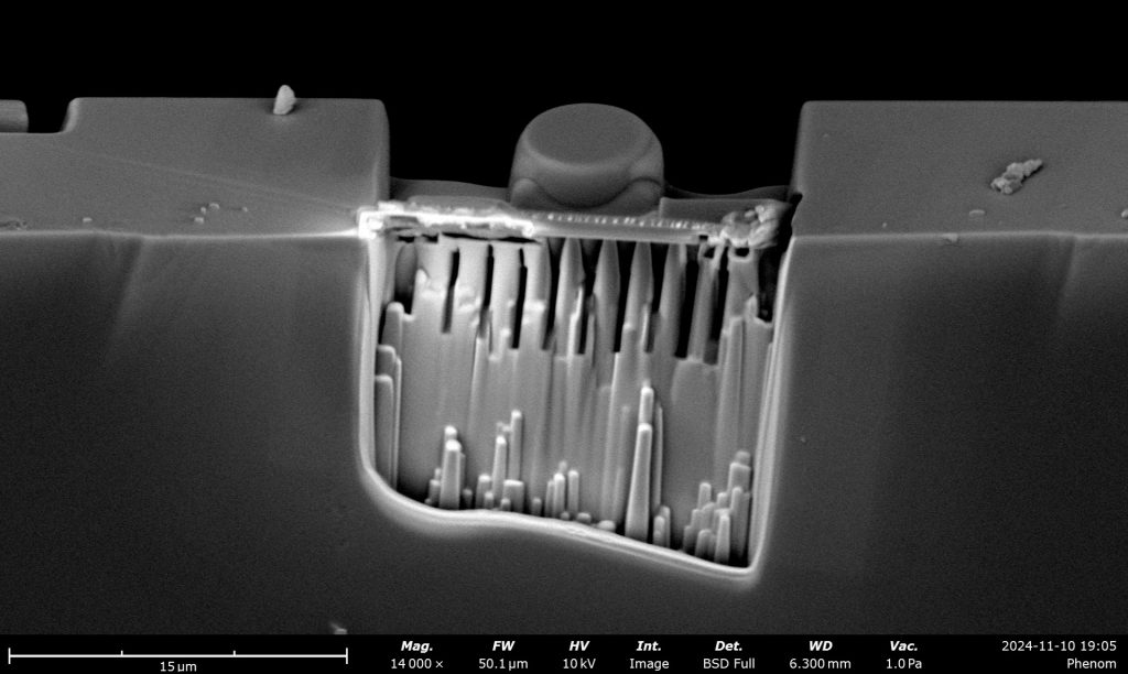
A micro MIT: A tiny replica of the Great Dome sculpted into silicon carbide using focused ion beam.
Alstroemeria anther with pollen.
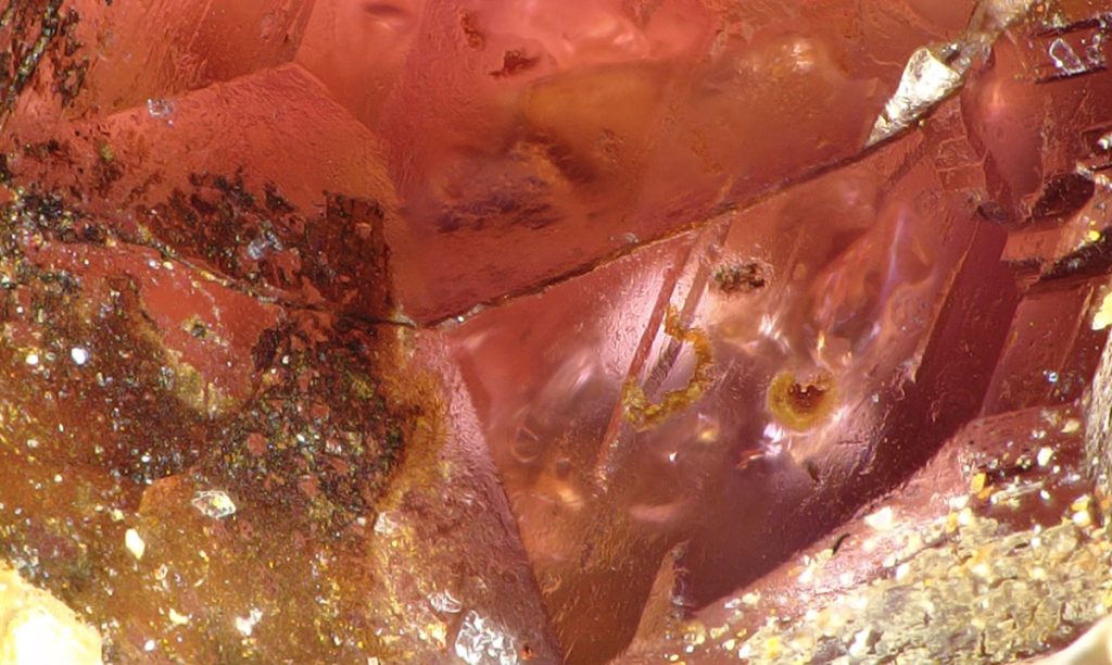
Garnet collected in Maine, showing its characteristic trapezohedral crystal habit—its natural crystal shape.
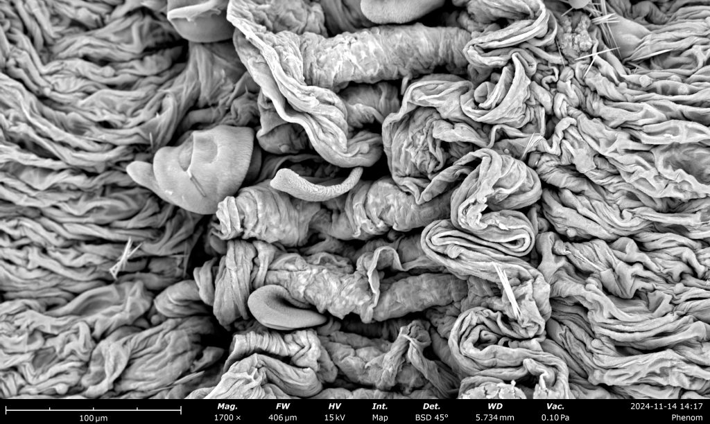
High-density polyethylene (HDPE) fibers from a torn event wristband.
Eye cells of a housefly.